42 human heart diagram with labels
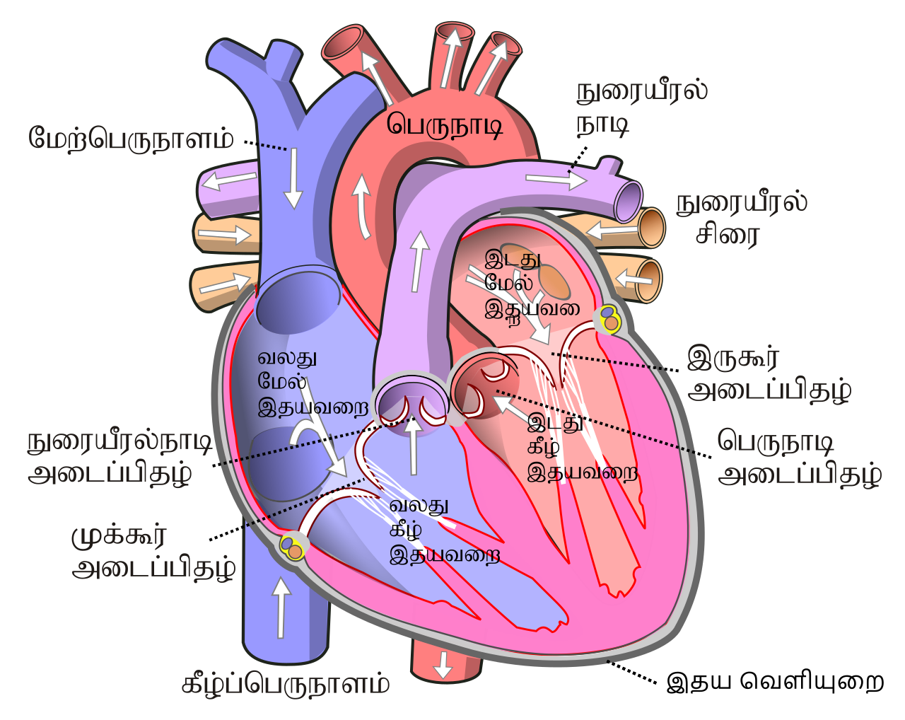
File:Diagram of the human heart (cropped).svg - Wikimedia Apr 05, 2022 · English: Diagram of the human heart 1. Superior vena cava 2. 4. Mitral valve 5. Aortic valve 6. Left ventricle 7. Right ventricle 8. Left atrium 9. Right atrium 10. Aorta 11. Pulmonary v The Human Heart Labeling Worksheet (Teacher-Made) - Twinkl The human heart is a muscle made up of four chambers, these are: Two lower chambers - the left and right ventricles. It's also made up of four valves - these are known as the tricuspid, pulmonary, mitral and aortic valves. With this heart diagram without labels, you can familiarise your students with all the correct terms and help them ...
A Diagram of the Heart and Its Functioning Explained in Detail The heart blood flow diagram (flowchart) given below will help you to understand the pathway of blood through the heart.Initial five points denotes impure or deoxygenated blood and the last five points denotes pure or oxygenated blood. 1.Different Parts of the Body ↓ 2.Major Veins ↓ 3.Right Atrium ↓ 4.Right Ventricle ↓ 5.Pulmonary Artery ↓ 6.Lungs

Human heart diagram with labels
Heart Diagram with Labels and Detailed Explanation The heart is located under the ribcage, between the lungs and above the diaphragm. It weighs about 10.5 ounces and is cone shaped in structure. It consists of the following parts: Heart Detailed Diagram Heart - Chambers There are four chambers of the heart . The upper two chambers are the auricles and the lower two are called ventricles. How to Draw a Human Heart: 11 Steps (with Pictures) - wikiHow 3. Sketch a forked tube extending from the top of the rounded bump. To make the superior vena cava, draw a tube coming from the top of the right atrium. Make the tube fork about the same length as the bump you made for the right atrium chamber. Blood enters the right atrium through the superior vena cava. Human Heart Drawing Tricks: Human Heart Diagram Class 10 Human Heart Diagram with Label In your exams, your diagram will be marked only if you have labeled it. That is how important labels are. If you practice the image multiple times with the labels, you will automatically remember all the marking labels. And to add to it, you must make sure all your marking labels are aligned.
Human heart diagram with labels. Human Heart (Anatomy): Diagram, Function, Chambers, Location in Body The heart is a muscular organ about the size of a fist, located just behind and slightly left of the breastbone. The heart pumps blood through the network of arteries and veins called the... Human Heart Diagram Pictures, Images and Stock Photos Human Heart Diagram Transparent blue image of human heart with monitored beat Antique illustration of heart Heart anatomy vector illustration. Labeled organ structure... Human heart anatomy, 3D illustration Abnormal ECG trace graph with a stethoscope Anatomy of Heart Anatomy of the human heart. Heart section. Blood flow. Heart Medical Exam Human Heart - Diagram and Anatomy of the Heart - Innerbody The heart is a muscular organ about the size of a closed fist that functions as the body's circulatory pump. It takes in deoxygenated blood through the veins and delivers it to the lungs for oxygenation before pumping it into the various arteries (which provide oxygen and nutrients to body tissues by transporting the blood throughout the body). Organ Map | Diagram of Human Body Internal Organs Functions Featuring an accurate illustration and informative, clear labels, this organ map is a fantastic visual aid to support your teaching during science lessons all about the human body's internal organs.Once downloaded, you'll have an A4 diagram of a human body and internal organs, clearly labelled and perfect for individual use. Each internal organ is labelled and includes a definition of each ...
Human Heart - Anatomy, Functions and Facts about Heart The human heart is one of the most important organs responsible for sustaining life. It is a muscular organ with four chambers. The size of the heart is the size of about a clenched fist. The human heart functions throughout a person’s lifespan and is one of the most robust and hardest working muscles in the human body. Human Heart Labeling Worksheets & Teaching Resources | TpT Human Heart Parts and Blood Flow Labeling Worksheets - Diagram/Graphic Organizer by TechCheck Lessons 14 $1.99 Zip This resource contains 2 worksheets for students to (1) label the parts of the human heart and (2) Fill in a flowchart tracing the path of blood flowing though the circulatory system. Answer keys included. 612 Human Heart Diagram Premium High Res Photos - Getty Images Browse 612 human heart diagram stock photos and images available, or search for heart illustration or pulmonary artery to find more great stock photos and pictures. An anatomical diagram showing the arteries of the human heart, circa 1930. Diagram of the human brain and other organs. File:Heart diagram-en.svg - Wikipedia File:Heart diagram-en.svg. Size of this PNG preview of this SVG file: 762 × 600 pixels. Other resolutions: 305 × 240 pixels | 610 × 480 pixels | 976 × 768 pixels | 1,280 × 1,008 pixels | 2,560 × 2,015 pixels | 893 × 703 pixels. This is a file from the Wikimedia Commons. Information from its description page there is shown below.
Human Heart Diagram - Human Body Pictures - Science for Kids Photo description: This is an excellent human heart diagram which uses different colors to show different parts and also labels a number of important heart component such as the aorta, pulmonary artery, pulmonary vein, left atrium, right atrium, left ventricle, right ventricle, inferior vena cava and superior vena cava among others. Male Human Anatomy Diagram Pictures, Images and Stock Photos Pacemaker Diagram Cross section of a human heart with pacemaker fitted, showing the major arteries and veins. This is an EPS 10 vector illustration and includes a high resolution JPEG. male human anatomy diagram stock illustrations Heart Diagram – 15+ Free Printable Word, Excel, EPS, PSD ... Teachers and students use the heart diagram, in biological science, to study the structure and functions of a human being’s heart. Friends and colleagues on the other hand may find this diagram template useful when it comes to sending special, personalized gifts to their family members and significant others. Download the template today, and ... Human Heart Diagram Without Labels - Pinterest The areas of the heart with MORE oxygen are labeled with an "R". Students will color these areas RED. The areas of the heart with LESS oxygen are labeled with a "B". Students will color these areas BLUE.
Heart Anatomy: Labeled Diagram, Structures, Function, and Blood Flow There are 4 chambers, labeled 1-4 on the diagram below. To help simplify things, we can convert the heart into a square. We will then divide that square into 4 different boxes which will represent the 4 chambers of the heart. The boxes are numbered to correlate with the labeled chambers on the cartoon diagram.
Human Heart Labeled Diagram The Human Heart Diagram Labeled - Human ... Jun 29, 2017 - Human Heart Labeled Diagram The Human Heart Diagram Labeled - Human Anatomy photo, Human Heart Labeled Diagram The Human Heart Diagram Labeled - Human Anatomy image, Human Heart Labeled Diagram The Human Heart Diagram Labeled - Human Anatomy gallery
Free Unlabelled Diagram Of The Heart, Download Free Unlabelled Diagram Of The Heart png images ...
A Labeled Diagram of the Human Heart You Really Need to See The human heart, comprises four chambers: right atrium, left atrium, right ventricle and left ventricle. The two upper chambers are called the left and the right atria, and the two lower chambers are known as the left and the right ventricles. The two atria and ventricles are separated from each other by a muscle wall called 'septum'.
Diagrams, quizzes and worksheets of the heart | Kenhub Worksheet showing unlabelled heart diagrams. Using our unlabeled heart diagrams, you can challenge yourself to identify the individual parts of the heart as indicated by the arrows and fill-in-the-blank spaces. This exercise will help you to identify your weak spots, so you'll know which heart structures you need to spend more time studying ...
Human Heart Diagram - Side View and Top View As shown in the human heart diagram above, the heart has several cavities (ventricles and atrium) and valves (pulmonary, aortic, mitral, tricuspid) which move blood in-and-out of the heart. It's pretty amazing to watch blood flow through the heart. To see how blood flows through the heart using an animation, please click here .
File:Diagram of the human heart (cropped).svg - Wikipedia File:Diagram of the human heart (cropped).svg. Size of this PNG preview of this SVG file: 611 × 600 pixels. Other resolutions: 244 × 240 pixels | 489 × 480 pixels | 782 × 768 pixels | 1,043 × 1,024 pixels | 2,086 × 2,048 pixels | 663 × 651 pixels. This is a file from the Wikimedia Commons. Information from its description page there is ...
BYJUS BYJUS
Human Heart Diagram Without Labels - Labelling Worksheet What makes up the anatomy of the human heart? The human heart is a muscle made up of four chambers, these are: Two upper chambers - the left atrium and right atrium Two lower chambers - the left and right ventricles. It's also made up of four valves - these are known as the tricuspid, pulmonary, mitral and aortic valves.
The Heart and Circulation of Blood - LSA The center of the circulatory system is the heart, which is the main pumping mechanism. The heart is made of muscle. The heart is shaped something like a cone, with a pointed bottom and a round top. It is hollow so that it can fill up with blood. An adult’s heart is about the size of a large orange and weighs a little less than a pound.

Human Heart Pictures with Labels Best Of File Diagram Of the Human Heart Hug Wikimedia Mons ...
File:Diagram of the human heart (no labels).svg - Wikimedia Size of this PNG preview of this SVG file: 498 × 599 pixels. Other resolutions: 199 × 240 pixels | 399 × 480 pixels | 499 × 600 pixels | 639 × 768 pixels | 851 × 1,024 pixels | 1,703 × 2,048 pixels | 533 × 641 pixels. Original file (SVG file, nominally 533 × 641 pixels, file size: 85 KB) File information. Structured data.

Abdominal Anatomy Male - Human Body Diagram Without Labels , Transparent Cartoon, Free Cliparts ...
Label the heart — Science Learning Hub Label the heart Add to collection In this interactive, you can label parts of the human heart. Drag and drop the text labels onto the boxes next to the diagram. Selecting or hovering over a box will highlight each area in the diagram. Pulmonary vein Right atrium Semilunar valve Left ventricle Vena cava Right ventricle Pulmonary artery Aorta

Heart Diagrams for Labeling and Coloring, With Reference Chart and Summary | Heart diagram ...
Human Heart Diagram Labeled | Science Trends Human Heart Diagram Labeled Daniel Nelson 1, January 2019 | Last Updated: 3, March 2020 The human heart is an organ responsible for pumping blood through the body, moving the blood (which carries valuable oxygen) to all the tissues in the body. Without the heart, the tissues couldn't get the oxygen they need and would die.
13+ Heart Diagram Templates - Sample, Example, Format Download Detail Human Heart Sample Template Response Sheet Of Blood And The Heart cfep.uci.edu This response sheet of blood and the heart contains all the parts of human heart including arteries, veins, atrium and so on. All the minute parts inside the human heart have been clearly labelled. Free Download Heart Structure And Its Functions Pdf Format
Heart Labeling Quiz: How Much You Know About Heart Labeling? Create your own Quiz Here is a Heart labeling quiz for you. The human heart is a vital organ for every human. The more healthy your heart is, the longer the chances you have of surviving, so you better take care of it. Take the following quiz to know how much you know about your heart. Questions and Answers 1. What is #1? 2. What is #2? 3.
Label Heart Anatomy Diagram Printout - EnchantedLearning.com Label Heart Interior Anatomy Diagram: Human Anatomy: The heart is a fist-sized, muscular organ that pumps blood through the body. Oxygen-poor blood enters the right atrium of the heart (via veins called the inferior vena cava and the superior vena cava). The blood is then pumped into the right ventricle and then through the pulmonary artery to ...
Human Heart Drawing Tricks: Human Heart Diagram Class 10 Human Heart Diagram with Label In your exams, your diagram will be marked only if you have labeled it. That is how important labels are. If you practice the image multiple times with the labels, you will automatically remember all the marking labels. And to add to it, you must make sure all your marking labels are aligned.
How to Draw a Human Heart: 11 Steps (with Pictures) - wikiHow 3. Sketch a forked tube extending from the top of the rounded bump. To make the superior vena cava, draw a tube coming from the top of the right atrium. Make the tube fork about the same length as the bump you made for the right atrium chamber. Blood enters the right atrium through the superior vena cava.


Post a Comment for "42 human heart diagram with labels"