41 fluorescent labels and light microscopy
Characterization of New Fluorescent Labels for Ultra-High ... Characterization of New Fluorescent Labels for Ultra-High Resolution Microscopy - PubMed Photo-induced switching of dyes into dark, long-lived states, such as a triplet state, has recently gained increasing interest, as a means to achieve ultra-high optical resolution. Label-free prediction of three-dimensional fluorescence images from ... Fluorescence microscopy can resolve subcellular structure in living cells, but is expensive, slow, and toxic. Here, we present a label-free method for predicting 3D fluorescence directly from transmitted light images and demonstrate its use to generate multi-structure, integrated images.
Fluorescence Microscopy - an overview | ScienceDirect Topics Fluorescence microscopy is a technique whereby fluorescent substances are examined in a microscope. It has a number of advantages over other forms of microscopy, offering high sensitivity and specificity. In fluorescence microscopy, the specimen is illuminated (excited) with light of a relatively short wavelength, usually blue or ultraviolet (UV).

Fluorescent labels and light microscopy
Fluorescent labeling of abundant reactive entities (FLARE) for ... - Nature Fluorescence microscopy is a technique that is commonly used in the biomedical sciences. It offers the powerful ability to visualize structures or molecules in three dimensions within biological... Imaging and Microscopy Fluorescence Filters - AZoOptics.com Fluorescence microscopes, imaging systems, flow cytometers, and DNA sequencers all use a light source, whether that is a laser, LED, or broadband lamp, in order to excite the fluorescent tags ... Seeing More with In Silico Labeling of Microscopy Images In contrast, fluorescence microscopy images are easier to analyze, because samples are prepared with carefully engineered fluorescent labels which light up just what the researchers want to see. For example, most human cells have exactly one nucleus, so a nuclear label (such as the blue one below) makes it possible for simple tools to find and ...
Fluorescent labels and light microscopy. Researchers demonstrate label-free super-resolution microscopy A newly developed sub-diffraction-limit microscopy approach doesn't require fluorescent labels. The video shows the process of the data evaluation algorithm, retrieving the positions and sizes of... A quick guide to light microscopy in cell biology - PMC Jan 15, 2016 · Fluorescence microscopy uses fluorescent dyes (fluorophores), which are molecules that absorb one wavelength of light (the excitation wavelength) and emit a second, longer wavelength of light (the emission wavelength). Fluorescent Dyes | Science Lab - Leica Microsystems In fluorescence microscopy there are two ways to visualize your protein of interest. Either with the help of an intrinsic fluorescent signal - by genetically linking a fluorescent protein to a target protein - or with the help of fluorescently labeled antibodies that bind specifically to a protein of interest. Fluorescence Microscopy - ChemBAM The technique of fluorescence microscopy has become an essential tool in biology and the biomedical sciences, as well as in materials science. The application of an array of fluorophores has made it possible to identify cells and sub-microscopic cellular components with a high degree of specificity (Fig. 3). These molecules can be conjugated to ...
Introduction to Fluorescence Microscopy | Nikon's MicroscopyU Introduction to Fluorescence Microscopy. The absorption and subsequent re-radiation of light by organic and inorganic specimens is typically the result of well-established physical phenomena described as being either fluorescence or phosphorescence. The emission of light through the fluorescence process is nearly simultaneous with the ... Fluorescent tag - Wikipedia Fluorescent labels can be hybridized to mRNA to help visualize interaction and activity, such as mRNA localization. An antisense strand labeled with the fluorescent probe is attached to a single mRNA strand, and can then be viewed during cell development to see the movement of mRNA within the cell. Fluorogenic labels Novel Fluorescent Label Shines a Light on DNA Structure in Cancer Cells Microscopy News Novel Fluorescent Label Shines a Light on DNA Structure in Cancer Cells March 7, 2022 0 Researchers have developed a new fluorescent label that gives a clearer picture of how DNA... Fluorescence Microscopy & Cell Imaging | Research | UNM Cancer Center Imaging. The Fluorescence Microscopy and Cell Imaging Shared Resource aids basic and physician researchers to image samples and publish high profile articles that: Elucidate cell and molecular mechanisms of cancer, immunologic, infectious, metabolic, neurologic and vascular diseases. Evaluate therapeutic efficacy in cells and patient samples.
Labeling the ER for Light and Fluorescence Microscopy Most of them are not 100% specific for the ER membrane and may label other organelles at varying concentrations and incubation times. ... C., Wang, P., Kriechbaumer, V. (2018). Labeling the ER for Light and Fluorescence Microscopy. In: Hawes, C., Kriechbaumer, V. (eds) The Plant Endoplasmic Reticulum . Methods in Molecular Biology, vol 1691 ... In Silico Labeling: Predicting Fluorescent Labels in Unlabeled ... - Cell The z-stacks of transmitted-light microscopy images were acquired with different methods for enhancing contrast in unlabeled images. Several different fluorescent labels were used to generate fluorescence images and were varied between training examples; the checkerboard images indicate fluorescent labels that were not acquired for a given example. Light Sheet Fluorescence Microscopy - an overview | ScienceDirect Topics Applications of single-molecule fluorescence microscopy. (A) The photophysical properties of a fluorophore contain information about its position and its state. This allows, for example, tracking molecules, observing conformational and constitutional changes, or following chemical reactions. (B) Examples for applications in biology and chemistry. Label-free prediction of three-dimensional fluorescence images from ... Label-free prediction of three-dimensional fluorescence images from transmitted-light microscopy Understanding cells as integrated systems is central to modern biology. Although fluorescence microscopy can resolve subcellular structure in living cells, it is expensive, is slow, and can damage cells.
Microscope World Blog: Fluorescence Microscopy
Light Microscope- Definition, Principle, Types, Parts, Labeled Diagram ... A light microscope is a biology laboratory instrument or tool, that uses visible light to detect and magnify very small objects and enlarge them. They use lenses to focus light on the specimen, magnifying it thus producing an image. The specimen is normally placed close to the microscopic lens.
Cell Preparation for Fixed Specimen Fluorescent Microscopy on Vimeo
Fluorescence Microscopy vs. Light Microscopy Nov 27, 2018 · This means that fluorescent microscopy uses reflected rather than transmitted light. For example, a commonly used label is green fluorescent protein (GFP), which is excited with blue light and...

Deltavision RT deconvolution fluorescence microscope (MCB) | The Microscopy Alliance | The ...
Fluorescence Imaging - Teledyne Photometrics Fluorescent molecules (known as fluorophores) are used to label samples, and fluorophores are available that emit light in virtually any color. In a fluorescent microscope, a sample is labeled with a fluorophore, and then a bright light ( excitation light) is used to illuminate the sample, which gives off fluorescence ( emission light ).

Light sheet fluorescence microscopy - YouTube
In Silico Labeling: Predicting Fluorescent Labels in Unlabeled Images Microscopy is a central method in life sciences. Many popular methods, such as antibody labeling, are used to add physical fluorescent labels to specific cellular constituents. ... (ISL), reliably predicts some fluorescent labels from transmitted-light images of unlabeled fixed or live biological samples. ISL predicts a range of labels, such as ...

Difference between Light Microscope and Electron Microscope (Light Microscope vs Electron ...
Light sheet fluorescence microscopy | Nature Reviews Methods Primers Light sheet fluorescence microscopy (LSFM) illuminates a thin slice of a fluorescent sample and observes the emission from this plane perpendicularly 1. In consequence, a light sheet microscope...

Three-photon microscopy improves biological imaging
Dots, Probes and Proteins: Fluorescent Labels for Microscopy and Imaging GFP now comes in 'flavors' including cyan, yellow and blue. Fluorescent proteins are useful for studying live cells and can be used as 'reporters' for studying gene expression. Using genetically modified plasmid and/or viral DNA, the target cells can be transfected with the plasmid which encodes both the fluorescent protein and a gene ...

A ‘super-resolution’ microscope for nanostructures | Kurzweil
Fluorescent Label - an overview | ScienceDirect Topics 14.3.2 Fluorescence quenching microscopy Fluorescence microscopy is a very common tool. Usually, fluorescent labels are used to brighten up the object of interest. However, the same strategy is not applicable for graphitic materials, such as graphite, graphene, GO or r-GO as they are strong quenchers of dye molecules [45].
.jpg)
Solutions for Spectral Imaging
Different Ways to Add Fluorescent Labels - Thermo Fisher Scientific Using fluorescence provides greater contrast compared to viewing your samples with brightfield microscopy alone. Labeling various targets with separate fluorescent colors allows you to visualize different structures or proteins within a cell in the same experiment.

Fluorescent Microscopy
Fluorescence Microscopy - Explanation and Labelled Images Dec 16, 2020 · Fluorescence microscopy uses a high-intensity light source that excites a fluorescent molecule called a fluorophore in the sample observed. The samples are labeled with fluorophore where they absorb the high-intensity light from the source and emit a lower energy light of longer wavelength.
Light-Sheet Fluorescence Microscopy Remote Training - France-BioImaging
Fluorescent Dyes in Microscopy - Types, Vs Proteins, Applications Etc. Fluorescent dyes (also known as fluorophores/reactive dyes) may simply be described as molecules (non-protein in nature) that, in microscopy, achieve their function by absorbing light at a given wavelength and re-emitting it at a longer wavelength. This produces fluorescence of different colors that can be visualized and analyzed.
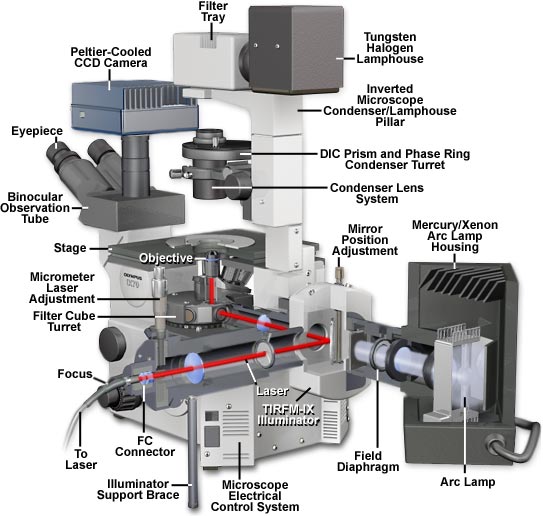
Molecular Expressions Microscopy Primer: Microscope Components - Olympus IX70 Microscope Cutaway ...
Different Ways to Add Fluorescent Labels - Thermo Fisher Using fluorescence provides greater contrast compared to viewing your samples with brightfield microscopy alone. Labeling various targets with separate ...
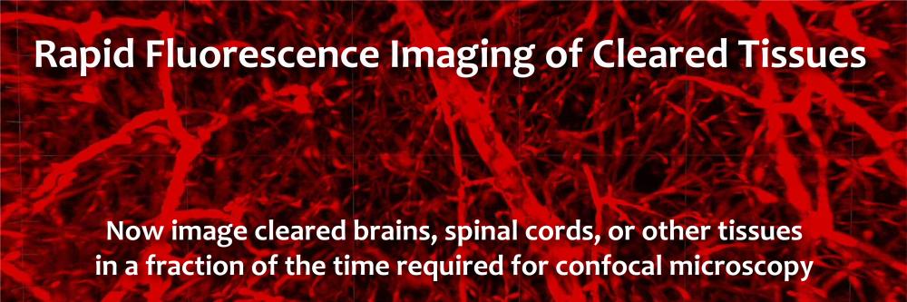
Light sheet fluorescence microscopy imaging of cleared tissues | Center - Imaging Center
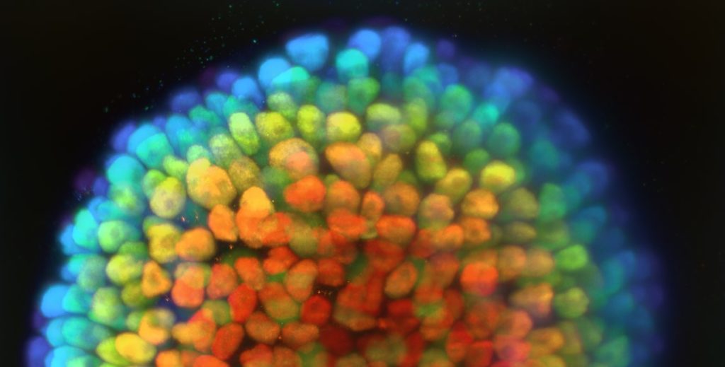
Imaging Flies by Fluorescence Microscopy: Principles, Technologies, and ... Fluorescence microscopy in combination with specific labeling methods [ e.g., antibodies or fluorescent proteins (FPs)] enables selective visualization of the components of living matter, from molecules and organelles to cells and tissues, in both fixed and living organisms, and with high signal-to-noise ratio (SNR).
News | CoolLED
Fluorescent Labeling - What You Should Know - PromoCell Fluorescence microscopy allows the identification of cells and cellular components and the monitoring of cell physiology with high specificity. Fluorescence microscopy separates emitted light from excitation light using optical filters. The use of two indicators also allows the simultaneous observation of different biomolecules at the same time.

Label-free prediction of three-dimensional fluorescence images from transmitted light microscopy ...
Seeing More with In Silico Labeling of Microscopy Images In contrast, fluorescence microscopy images are easier to analyze, because samples are prepared with carefully engineered fluorescent labels which light up just what the researchers want to see. For example, most human cells have exactly one nucleus, so a nuclear label (such as the blue one below) makes it possible for simple tools to find and ...

Label-free prediction of three-dimensional fluorescence images from transmitted-light microscopy ...
Imaging and Microscopy Fluorescence Filters - AZoOptics.com Fluorescence microscopes, imaging systems, flow cytometers, and DNA sequencers all use a light source, whether that is a laser, LED, or broadband lamp, in order to excite the fluorescent tags ...

New Technologies at Neuroscience 2012 | Biocompare Editorial Article
Fluorescent labeling of abundant reactive entities (FLARE) for ... - Nature Fluorescence microscopy is a technique that is commonly used in the biomedical sciences. It offers the powerful ability to visualize structures or molecules in three dimensions within biological...

Microbiology Notes: The Study of Microbial Structure : Microscopy and Specimen Preparation
Post a Comment for "41 fluorescent labels and light microscopy"