43 microscope diagram without labels
Compound Microscope Parts - Labeled Diagram and their Functions The eyepiece (or ocular lens) is the lens part at the top of a microscope that the viewer looks through. The standard eyepiece has a magnification of 10x. You may exchange with an optional eyepiece ranging from 5x - 30x. [In this figure] The structure inside an eyepiece. The current design of the eyepiece is no longer a single convex lens. 7th grade Science - Microscope Diagram | Quizlet The Parts of a Microscope. 12 terms. totobear PLUS. Sets found in the same folder. Science Key terms 7th grade. 13 terms. palocastillo. 7th Grade Earth Science. 9 terms. EliseC17. 7thGrade Review - Cells/Biology. 26 terms. SolizScience TEACHER. 7th grade Science, Cell theory. 8 terms. Super1412. Other sets by this creator.
Microscope Diagram - cell division of e coli with continuous media flow ... Microscope Diagram - 15 images - give a well labelled diagram of compound microscope using of typical, bio tem biological transmission electron microscope university, labelled microscope diagram gcse micropedia, a compound microscope diagram micropedia,

Microscope diagram without labels
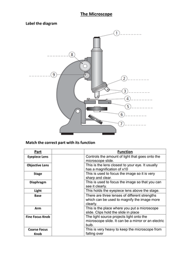
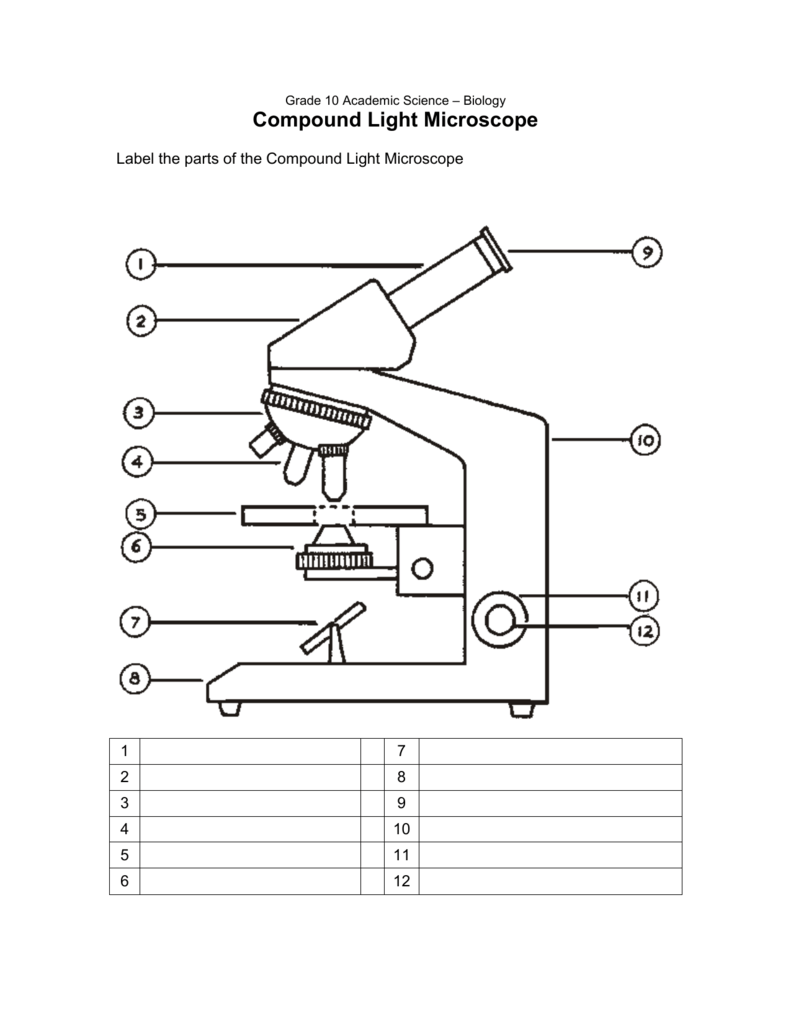
16 Parts of a Compound Microscope: Diagrams and Video Once you have an understanding of the parts of the microscope it will be much easier to navigate around and begin observing your specimen, which is the fun part! The 16 core parts of a compound microscope are: Head (Body) Arm Base Eyepiece Eyepiece tube Objective lenses Revolving Nosepiece (Turret) Rack stop Coarse adjustment knobs Free Microscope Worksheets for Simple Science Fun for Your Students 1. Parts of a Microscope . The first worksheet labels the different parts of a microscope, including the base, slide holder, and condenser. If you have a microscope, compare and contrast this worksheet to it.Also, your kids can color this microscope diagram in and read the words to each part of the microscope. Microscope Parts and Functions First, the purpose of a microscope is to magnify a small object or to magnify the fine details of a larger object in order to examine minute specimens that cannot be seen by the naked eye. Here are the important compound microscope parts... Eyepiece: The lens the viewer looks through to see the specimen.
Microscope diagram without labels. Compound Microscope: Definition, Diagram, Parts, Uses, Working ... - BYJUS A compound microscope is defined as. A microscope with a high resolution and uses two sets of lenses providing a 2-dimensional image of the sample. The term compound refers to the usage of more than one lens in the microscope. Also, the compound microscope is one of the types of optical microscopes. The other type of optical microscope is a ... Microscope, Microscope Parts, Labeled Diagram, and Functions The Iris Diaphragm is located above the condenser lens and below the microscope stage. The different sized holes in the diaphragm helps to vary the size of the cone and intensity of light that is projected upward into the slide. However, there is no set rule regarding which setting to use for a particular power. Labelled Diagram of Compound Microscope - Biology Discussion The below mentioned article provides a labelled diagram of compound microscope. Part # 1. The Stand: The stand is made up of a heavy foot which carries a curved inclinable limb or arm bearing the body tube. The foot is generally horse shoe-shaped structure (Fig. 2) which rests on table top or any other surface on which the microscope in kept. pages.zeiss.com › rs › 896-XMS-794Principles of Fluorescence and Fluorescence Microscopy - ZEISS fies the principle of the fluorescence microscope — without the light-filtering abilities of the purple glass window and the glass of white wine, Stokes would not have been able to observe any fluorescence at all. Using Stokes’ observation and the green fluorescent protein (GFP) as examples, this article will explain
en.wikipedia.org › wiki › FluorescenceFluorescence - Wikipedia Fluorescence is the emission of light by a substance that has absorbed light or other electromagnetic radiation.It is a form of luminescence.In most cases, the emitted light has a longer wavelength, and therefore a lower photon energy, than the absorbed radiation. A Study of the Microscope and its Functions With a Labeled Diagram ... Here, unlabeled microscope diagrams have been provided for your perusal, which will help you practice and test your understanding of the instrument. Types of Microscopes Depending on the source of illumination, microscopes can be divided into two categories. They are: › books › NBK26880Looking at the Structure of Cells in the Microscope ... A special sample holder is used to keep this hydrated specimen at -160°C in the vacuum of the microscope, where it can be viewed directly without fixation, staining, or drying. Unlike negative staining, in which what is seen is the envelope of stain exclusion around the particle, hydrated cryoelectron microscopy produces an image from the ... › products › microscopeMicroscope Objective Lens | Products | Leica Microsystems The objective lens is a critical part of the microscope optics. The microscope objective is positioned near the sample, specimen, or object being observed. It has a very important role in imaging, as it forms the first magnified image of the sample. The numerical aperture (NA) of the objective indicates its ability to gather light and largely determines the microscope’s resolution, the ...
Label A Microscope Teaching Resources | Teachers Pay Teachers The 13 parts of the microscope: microscope, base, arm, inclination joint, course adjustment, fine adjustment, body tube, ocular lens, revolving nose piece, objectives, stage, stage clips, and iris diaphragm.Includes:13 cards with labels13 cards without labels13 labels1 blackline masterCards with labels are approx. 3¾" x 4", cards without ... PDF Parts of a Microscope Printables - Homeschool Creations Label the parts of the microscope. You can use the word bank below to fill in the blanks or cut and paste the words at the bottom. ... without needing to move the microscope ? the head •What is the magnification level on the eyepiece of a microscope?10x (see objective blank cell diagram to label microscope diagram unlabeled light parts blank biology science labeled lab sketch worksheet labels worksheets timvandevall drawing quiz chemistry microscopes fill Mitosis Diagram Without Labels For Kids - Simple Animal Cell Unlabelled peroxisome Cell Diagram To Label - Pensandpieces pensandpieces.blogspot.com ixl organelle Diagram of a Compound Microscope - Biology Discussion 1. It is noted first that which objective lens is in use on the microscope. 2. Stage micrometer is positioned in such a way that it is in the field of view. 3. The eyepiece is rotated so that the two scales, the eyepiece or ocular scale and the stage micrometer scale, are parallel. 4.
All Saints Online
Parts of the Microscope with Labeling (also Free Printouts) A microscope is one of the invaluable tools in the laboratory setting. It is used to observe things that cannot be seen by the naked eye. Table of Contents 1. Eyepiece 2. Body tube/Head 3. Turret/Nose piece 4. Objective lenses 5. Knobs (fine and coarse) 6. Stage and stage clips 7. Aperture 9. Condenser 10. Condenser focus knob 11. Iris diaphragm

give a well labelled diagram of compound microscope using of typical animal as well as plant ...
› en › microscopeFluorescence Resonance Energy Transfer (FRET) Microscopy Presented in Figure 3 is a Jablonski diagram illustrating the coupled transitions involved between the donor emission and acceptor absorbance in fluorescence resonance energy transfer. Absorption and emission transitions are represented by straight vertical arrows (green and red, respectively), while vibrational relaxation is indicated by wavy ...
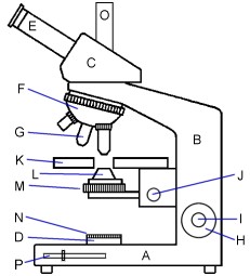
Diagram Of A Microscope With Labels - Drivenhelios
Compound Microscope Parts, Functions, and Labeled Diagram Compound Microscope Definitions for Labels. Eyepiece (ocular lens) with or without Pointer: The part that is looked through at the top of the compound microscope. Eyepieces typically have a magnification between 5x & 30x. Monocular or Binocular Head: Structural support that holds & connects the eyepieces to the objective lenses.

Search in gallery
en.wikipedia.org › wiki › Electron_microscopeElectron microscope - Wikipedia An electron microscope is a microscope that uses a beam of accelerated electrons as a source of illumination. As the wavelength of an electron can be up to 100,000 times shorter than that of visible light photons, electron microscopes have a higher resolving power than light microscopes and can reveal the structure of smaller objects.
Labelled Microscope Diagram Gcse - Micropedia
› confocal-microscopes › lsm-980LSM 980 with Airyscan 2 – Confocal Microscope with Multiplex ... This requires excellent imaging performance combined with low phototoxicity and high speed. LSM 980, your platform for confocal 4D imaging, is optimized for simultaneous spectral detection of multiple weak labels with the highest light efficiency. Employ a wealth of fluorescent labels from 380 nm to 900 nm.

Microscope World Blog: High School Microscope Features
Label the microscope — Science Learning Hub In this interactive, you can label the different parts of a microscope. Use this with the Microscope parts activity to help students identify and label the main parts of a microscope and then describe their functions. Drag and drop the text labels onto the microscope diagram.

8 Best Images of Lens Diagram Worksheet - Microscope with Labeled Parts, Label Eye Parts ...
Microscope Label Interactive Worksheets & Teaching Resources | TpT Microscope Interactive Notebook Activity by Jodi's Jewels 12 $1.89 PDF Students will complete a timeline of the history of the microscope, label a diagram, and create a pocket foldable with terms and definition cards. The timeline can be completed according to the teacher's directions or like the answer key example.

Cell Organelles | Cells: The Basic Units Of Life | Siyavula
Label the Microscope Diagram | Download Scientific Diagram - ResearchGate Download scientific diagram | Label the Microscope Diagram from publication: Laboratory Exercises in Microbiology: Discovering the Unseen World through Hands-on Investigation | Microbiology ...

www.timvandevall.com wp-content uploads Labeled-Microscope-Diagram.jpg | ชีววิทยาศาสตร์, ห้อง ...
Parts of Stereo Microscope (Dissecting microscope) - labeled diagram ... Stereo microscopes (also called Dissecting microscope) are branched out from other light microscopes for the application of viewing "3D" objects. These include substantial specimens, such as insects, feathers, leaves, rocks, sand grains, gems, coins, and stamps, etc. Functionally, a stereo microscope is like a powerful magnifying glass.

Label Microscope Diagram - ClipArt Best
Parts of a microscope with functions and labeled diagram - Microbe Notes Figure: Diagram of parts of a microscope There are three structural parts of the microscope i.e. head, base, and arm. Head - This is also known as the body. It carries the optical parts in the upper part of the microscope. Base - It acts as microscopes support. It also carries microscopic illuminators.

Chapter 1 Questions PPT - BIOLOGY JUNCTION
Label Microscope Diagram - EnchantedLearning.com Using the terms listed below, label the microscope diagram. arm - this attaches the eyepiece and body tube to the base. base - this supports the microscope. body tube - the tube that supports the eyepiece. coarse focus adjustment - a knob that makes large adjustments to the focus. diaphragm - an adjustable opening under the stage, allowing ...
Microscope Labelled Diagram Gcse - Micropedia
PDF Label compound microscope worksheet - Weebly [clearBoth] [clearBoth] Microscope diagram without label After you've studied all the pieces of the composite microscope, it's time to put your brain to the test. Print an unmarked microscope chart and check that you can fill out all the blanks. [clearBoth] [clearBoth] Blank microscope diagram Next we have an empty microscope diagram.

The Wonderful Microworld: Cell Nucleus - Onion
Simple Microscope - Parts, Functions, Diagram and Labelling A simple microscope is a device that only has one lens for magnification. It functions the same way as the magnifying glass. Although it is simple in terms of design and function, it is useful I various fields including medicine, jewelry and watchmaking, and agriculture, to name a few. References

Kenya Forensics Online Resource: CARDIAC MUSCLE TISSUE
PDF Label parts of the Microscope: Answers Label parts of the Microscope: Answers Coarse Focus Fine Focus Eyepiece Arm Rack Stop Stage Clip . Created Date: 20150715115425Z ...
33 Microscope Diagram To Label - Labels Database 2020
Microscope Labeling - The Biology Corner Students label the parts of the microscope in this photo of a basic laboratory light microscope. Can be used for practice or as a quiz. ... The type of microscope used in most science classes is the _____ microscope. 18. You should carry the microscope by the _____ and the _____. 19. The objectives are attached to what part of the microscope ...

Draw the cell of onion peel as seen under Microscope. state their features - Brainly.in
Amazing 27 Things Under The Microscope With Diagrams - Microbe Notes Under a high-power microscope, the cell organelles are more differentiated and allow the observation of individual structures. Because of the affinity of the stain with the DNA and RNA of the cell, the components inside the nucleus might also be visible. 10. DNA under the microscope.

Marchantia polymorpha. Common liverwort. Archegoniophore. Longitudinal section. 64X - Sporophyte ...
How does a Microscope work A compound microscope has two or more lenses. The eyepiece or ocular lens sits atop the body tube. Many microscopes are binocular and have two ocular lenses. Additionally, a binocular head will have a prism, either in the head or the body tube, to split the image and direct it to both oculars.
Post a Comment for "43 microscope diagram without labels"