44 external structure of the heart with labels
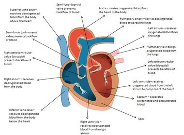
Chapter 22 Heart Flashcards | Quizlet Label the coronary arteries in an anterior view of the heart. Label the order that blood flows through in the heart, using the arrows as guides. Label the components of the heart wall. Label the components of the heart as seen from a posterior view. Label the major coronary veins. Label the components of the conduction system. Human Heart - Diagram and Anatomy of the Heart - Innerbody Because the heart points to the left, about 2/3 of the heart's mass is found on the left side of the body and the other 1/3 is on the right. Anatomy of the Heart Pericardium. The heart sits within a fluid-filled cavity called the pericardial cavity. The walls and lining of the pericardial cavity are a special membrane known as the pericardium.
Solved Help Label the external anatomy on this posterior - Chegg Question: Help Label the external anatomy on this posterior view of a mammalian heart by clicking and dragging the labels to the correct location Coronary sinus Apex of heart Lert atrium Posterior interventricular branch of LCA Left pulmonary artery Left ventricle Left pulmonary veins Aortic arch This problem has been solved! See the answer

External structure of the heart with labels
Heart Anatomy: size, location, coverings and layers : Anatomy & Physiology Heart Anatomy. The heart is around the size of a fist and weighs between 250-350 grams (less than a pound). Enclosed within the mediastinum, the medial cavity of the thorax, the heart extends obliquely from the second rib to the fifth intercostal space. It rests on the superior surface of the diaphragm, lies posterior to the sternum and ... Heart Labeling Quiz: How Much You Know About Heart Labeling? Here is a Heart labeling quiz for you. The human heart is a vital organ for every human. The more healthy your heart is, the longer the chances you have of surviving, so you better take care of it. Take the following quiz to know how much you know about your heart. Questions and Answers 1. What is #1? 2. What is #2? 3. What is #3? 4. What is #4? 19.1 Heart Anatomy - Anatomy and Physiology 2e | OpenStax Location of the Heart. The human heart is located within the thoracic cavity, medially between the lungs in the space known as the mediastinum. Figure 19.2 shows the position of the heart within the thoracic cavity. Within the mediastinum, the heart is separated from the other mediastinal structures by a tough membrane known as the pericardium, or pericardial sac, and sits in its own space ...
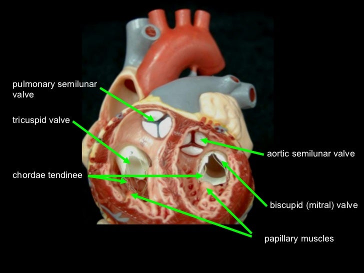
External structure of the heart with labels. Label the heart — Science Learning Hub Label the heart Interactive Add to collection In this interactive, you can label parts of the human heart. Drag and drop the text labels onto the boxes next to the diagram. Selecting or hovering over a box will highlight each area in the diagram. pulmonary vein semilunar valve right ventricle right atrium vena cava left atrium pulmonary artery Heart - External Features - Anatomy QA Apex beat. Is the lowermost and outermost thrust of the heart, felt on the front of the chest. In adults it is felt in the left 5 th intercostal space 9cm. from the median plane (just medial to the midclavicular line). In infants it is felt in the 3 rd intercostal space just lateral to the midclavicular line.. Dextrocardia. It is a congenital anomaly in which the heart lies on the right side ... Heart anatomy: Structure, valves, coronary vessels | Kenhub The heart is shaped as a quadrangular pyramid, and orientated as if the pyramid has fallen onto one of its sides so that its base faces the posterior thoracic wall, and its apex is pointed toward the anterior thoracic wall. Internal Structure of the Heart | Contemporary Health Issues It is marked by the presence of four openings that allow blood to move from the atria into the ventricles and from the ventricles into the pulmonary trunk and aorta. Located in each of these openings between the atria and ventricles is a valve, a specialized structure that ensures one-way flow of blood.
Heart Anatomy | Anatomy and Physiology II - Lumen Learning The wall of the heart is composed of three layers of unequal thickness. From superficial to deep, these are the epicardium, the myocardium, and the endocardium. The outermost layer of the wall of the heart is also the innermost layer of the pericardium, the epicardium, or the visceral pericardium discussed earlier. Figure 6. Heart Diagram with Labels and Detailed Explanation - BYJUS Diagram of Heart. The human heart is the most crucial organ of the human body. It pumps blood from the heart to different parts of the body and back to the heart. The most common heart attack symptoms or warning signs are chest pain, breathlessness, nausea, sweating etc. The diagram of heart is beneficial for Class 10 and 12 and is frequently ... The Anatomy of the Heart, Its Structures, and Functions - ThoughtCo The heart is the organ that helps supply blood and oxygen to all parts of the body. It is divided by a partition (or septum) into two halves. The halves are, in turn, divided into four chambers. The heart is situated within the chest cavity and surrounded by a fluid-filled sac called the pericardium. This amazing muscle produces electrical ... Layers of the heart: Epicardium, myocardium, endocardium - Kenhub The endocardium is the innermost layer of the heart. It lines the inner surfaces of the heart chambers, including the heart valves. The endocardium has two layers. The inner layer lines the heart chambers and is made of endothelial cells.
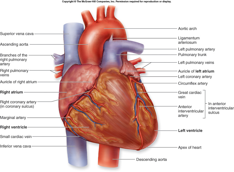
Solved Art-Labeling Activity: Overview of the external - Chegg art-labeling activity: overview of the external anatomy of the heart anterior view res great cardiac vein aortic arch right coronary artery left coronary artery left pulmonary veins ascending aorta left pulmonary artery anterior interventricular artery superior vena cava pulmonary trunk auricle of left atrium circumflex artery auricle of right … How to Draw the Internal Structure of the Heart (with Pictures) - wikiHow To draw the internal structure of a human heart, follow the steps below. Part 1 Finding a Diagram 1 To find a good diagram, go to Google Images, and type in "The Internal Structure of the Human Heart". Find an image that displays the entire heart, and click on it to enlarge it. 2 Find a piece of paper and something to draw with. Correctly Label The Following External Anatomy Of The Anterior Heart ... The external anatomy of the human heart consists of the four chambers that form the apex of the heart. Each chamber has an apex that corresponds to a box. There are two boxes on each side of the heart: the atria and the ventricles. The left atrium is a branching organ. The pulmonary trunk contains the aorta and pulmonary veins. Heart Anatomy Labeling Game - PurposeGames.com This is an online quiz called Heart Anatomy Labeling Game. There is a printable worksheet available for download here so you can take the quiz with pen and paper. Your Skills & Rank. Total Points. 0. Get started! Today's Rank--0. Today 's Points. One of us! Game Points. 19. You need to get 100% to score the 19 points available.
Human Bio 156: Compendium Review Unit 2 Major Topic: Oxygen/Microbes/Immunity
Label the Heart - The Biology Corner A simple heart diagram with arrows and boxes for students to practice labeling the chambers and major vessels. Name:_____Date: _____ Label the Heart. Word Bank: Left Atrium | Right Atrium | Left Ventricle | Right Ventricle Aorta | Pulmonary Veins | Pulmonary Artery | Superior Vena Cava | Inferior Vena Cava Bicuspid Valve | Tricuspid Valve ...
Structure and Function of the Heart
Structure of the Heart | SEER Training - National Cancer Institute The human heart is a four-chambered muscular organ, shaped and sized roughly like a man's closed fist with two-thirds of the mass to the left of midline. The heart is enclosed in a pericardial sac that is lined with the parietal layers of a serous membrane. The visceral layer of the serous membrane forms the epicardium. Layers of the Heart Wall

Heart Diagram Labeled Posterior - Diagram Media
External anterior heart labeling Quiz - purposegames.com About this Quiz This is an online quiz called External anterior heart labeling There is a printable worksheet available for download here so you can take the quiz with pen and paper. Your Skills & Rank Total Points 0 Get started! Today's Rank -- 0 Today 's Points One of us! Game Points 27 You need to get 100% to score the 27 points available

Fig 2 Gross Anatomy of the Heart (c)
Diagram of the human heart royalty-free images - Shutterstock Find Diagram of the human heart stock images in HD and millions of other royalty-free stock photos, illustrations and vectors in the Shutterstock collection. Thousands of new, high-quality pictures added every day.

Structure Of The Heart | A-Level Biology Revision Notes The heart is a hollow muscular organ that lies in the middle of the chest cavity. It is enclosed in the pericardium, which protects the heart and facilitates its pumping action. The heart is divided into four chambers: The two atria (auricles): these are the upper two chambers. They have thin walls which receive blood from veins.
Heart without labels
2. External features of the heart - SlideShare Chambers of the Heart • The heart is divided by 2 septa (interatrial and interventricular septa) into four chambers: 1. The right and left atria 2. The right and left ventricles. • The right atrium lies anterior to the left atrium, and the right ventricle lies anterior to the left ventricle 12.
Free Blank Heart Diagram, Download Free Blank Heart Diagram png images, Free ClipArts on Clipart ...
A Labeled Diagram of the Human Heart You Really Need to See The human heart, comprises four chambers: right atrium, left atrium, right ventricle and left ventricle. The two upper chambers are called the left and the right atria, and the two lower chambers are known as the left and the right ventricles. The two atria and ventricles are separated from each other by a muscle wall called 'septum'.
The structure of the heart | Slide Set
Heart Anatomy: Heart Dissection - University of Washington The major vessels of the heart are found at the base of the heart, along with the upper chambers, the right atrium (C) and left atrium (D). The atria are collapsed, but in a functioning heart, they would be stretched full of blood. The majority of the heart tissue consists of the ventricles. The left ventricle (F) is stiff and solid because it ...
Activity 9-blood-heart
Label the Heart - The Biology Corner Shows a picture of a heart with letters and blanks for practice with labeling the parts of the heart and tracing the flow of blood within the heart.

Ch. 19 Circulatory System- heart Flashcards | Quizlet Correctly label the external anatomy of the anterior heart. Place the labels in order denoting the flow of blood through the pulmonary circuit beginning with the right atrium and ending in the left atrioventricular valve. The first and last structures are given. Right atrium 1. tricuspid valve 2. right ventricle 3. pulmonary valve
The Heart - Labelled diagram
External Heart Anatomy labeled.jpg - B. External Anatomy of... View External Heart Anatomy labeled.jpg from BIOL 186 at Messiah. B. External Anatomy of the heart (label diagram, Figure 17.5, p636-637) blue - path of oxygen poor blood arteries away from red- path

HR & BP - bright's blogs
19.1 Heart Anatomy - Anatomy and Physiology 2e | OpenStax Location of the Heart. The human heart is located within the thoracic cavity, medially between the lungs in the space known as the mediastinum. Figure 19.2 shows the position of the heart within the thoracic cavity. Within the mediastinum, the heart is separated from the other mediastinal structures by a tough membrane known as the pericardium, or pericardial sac, and sits in its own space ...

Important images for system of frog
Heart Labeling Quiz: How Much You Know About Heart Labeling? Here is a Heart labeling quiz for you. The human heart is a vital organ for every human. The more healthy your heart is, the longer the chances you have of surviving, so you better take care of it. Take the following quiz to know how much you know about your heart. Questions and Answers 1. What is #1? 2. What is #2? 3. What is #3? 4. What is #4?

Eye Makeup Diagram for Eyeliner Application - Maybelline
Heart Anatomy: size, location, coverings and layers : Anatomy & Physiology Heart Anatomy. The heart is around the size of a fist and weighs between 250-350 grams (less than a pound). Enclosed within the mediastinum, the medial cavity of the thorax, the heart extends obliquely from the second rib to the fifth intercostal space. It rests on the superior surface of the diaphragm, lies posterior to the sternum and ...
heart - Cmap for Heart Internal view Cardiac Output

External Structure Of The Heart | biology // physiology | Pinterest
Post a Comment for "44 external structure of the heart with labels"